Cross Section Of A Compact Bone - 1 Oral embryology, histology and anatomy | Pocket Dentistry : These are abundant and characteristic of compact bone.. A cross section of a long bone, showing the internal structure. This model shows a cross section of compact bone. Compact bone, makes up the dense material in a long section of a bone. Cross section of compact bone. These are abundant and characteristic of compact bone.
Observe that the matrix of the bone is deposited in concentric layers that are called lamellae (5). Bone decalcification is the removal of the mineral component using an acid, leaving the bone soft and easy to cut. The two layers of compact bone and the interior spongy bone work together to protect the internal organs. Also called cortical bone, the compact variety usually features a haversian system, or cylindrical unit within the structure. These are abundant and characteristic of compact bone.
Compact bone - Stock Image - P105/0168 - Science Photo Library from media.sciencephoto.com Compact bone & spongy bone. Compact bone, makes up the dense material in a long section of a bone. Osteocyte processes lie in tiny canals (canaliculi) in the bone matrix. Don't assume that the cross sectional area is the same no matter where you cut. Most bones contain both compact and spongy bone. Observe that the matrix of the bone is deposited in concentric layers that are called lamellae (5). A cross section of a human long bone. The basic unit of structure in this type of bone is the haversian system, or osteon.
Most bones contain both compact and spongy bone.
Also called cortical bone, the compact variety usually features a haversian system, or cylindrical unit within the structure. There are trabeculae in spongy bone which gives its sponge like appearance. Compact bone, also known as cortical bone, is a denser material used to create much of the hard structure of the skeleton. Canaliculi allow the passage of interstitial fluid between the central canal and the lacunae housing osteocytes. Hope you enjoy and please. Cross section of compact bone. Bone decalcification is the removal of the mineral component using an acid, leaving the bone soft and easy to cut. In this sample, both compact, cortical bone as well as some mature. This is a cross section through decalcified bone. Between the rings of matrix, the bone cells (osteocytes) are located in spaces called lacunae. The remainder is spongelike cancellous bone. Structures and bone areas in column b, and use them to color the coding. Compact bone consists of closely packed osteons or haversian systems.
Concentric layers of bone cells (osteocytes). Details available from source webpage. Also called cortical bone, the compact variety usually features a haversian system, or cylindrical unit within the structure. A diagrammatic view of a cross section of bone. These are abundant and characteristic of compact bone.
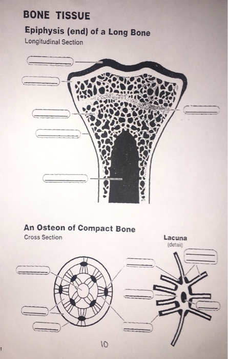
Solved: BONE TISSUE Epiphysis (end) Of A Long Bone Longitu ... from media.cheggcdn.com Compact bone, makes up the dense material in a long section of a bone. Cross section of compact bone. A cross section of a long bone, showing the internal structure. As seen in the image below each osteon is also composed of a number of different cells responsible for the maintenance of the bones, including osteocytes and osteoblasts. Compact bone consists of closely packed osteons or haversian systems. As the names suggest compact bone looks compact and the spongy bone looks like sponges. The remaining material is mostly collagen. A cross section of a human long bone.
Cross section of compact bone.
Cross section of compact bone. The two layers of compact bone and the interior spongy bone work together to protect the internal organs. Their course follows the main axis of long bone. Hope you enjoy and please. (b) in this micrograph of the osteon, you can clearly see the concentric lamellae and central canals. (micrograph provided by the regents of university of michigan. These are abundant and characteristic of compact bone. This model shows a cross section of compact bone. The outlined area is a cross section of an osteon of compact bone. Cross section of the compact bone. Select different colors for the. Compact bone & spongy bone. In each of these osteons, the lamellae are arranged around a central.
The outlined area is a cross section of an osteon of compact bone. This model shows a cross section of compact bone. They build the entire picture, improve your understanding, consolidate the information and facilitate recall. Spongy bone is the osseous tissue, which fills the interior cavity of bones, consisting of mineralized bars called trabeculae. The outlined area is a cross section of an osteon of compact bone.
Histology > Garland > Flashcards > 5- Bone | StudyBlue from classconnection.s3.amazonaws.com They build the entire picture, improve your understanding, consolidate the information and facilitate recall. Also called cortical bone, the compact variety usually features a haversian system, or cylindrical unit within the structure. A cross section of a compact bone shows concentric circles called lamellae. Compact bone & spongy bone. Don't assume that the cross sectional area is the same no matter where you cut. In three dimensions an osteon is cylindrical in shape. Bone must be decalcified (by exposure to strong acids) so it can be cut into thin sections. The two layers of compact bone and the interior spongy bone work together to protect the internal organs.
Canaliculi allow the passage of interstitial fluid between the central canal and the lacunae housing osteocytes.
Compact bone consists of closely packed osteons or haversian systems. This model shows a cross section of compact bone. Cross section of compact bone. Don't assume that the cross sectional area is the same no matter where you cut. There are two ways to study bone histology. Between the rings of matrix, the bone cells (osteocytes) are located in spaces called lacunae. Most bones contain both compact and spongy bone. The remainder is spongelike cancellous bone. Compact bone, also known as cortical bone, is a denser material used to create much of the hard structure of the skeleton. This is a short tutorial using blender 2.8 that shows how to create a bone cross section and using images to create the textures. The basic unit of structure in this type of bone is the haversian system, or osteon. Bone must be decalcified (by exposure to strong acids) so it can be cut into thin sections. The connection point for the periosteum.
A cross section of a long bone, showing the internal structure cross section of a bone. Cross section of the compact bone.


0 Komentar